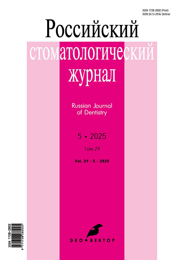
Russian Journal of Dentistry
Peer-review bimonthly medical journal.
Editor-in-chief
- Valentina N. Olesova, MD, Dr. Sci. (Medicine), Professor
ORCID iD: 0000-0002-3461-9317
Publisher
- Eco-Vector Publishing Group
WEB: https://eco-vector.com/
About
The journal covers relevant issues in dentistry, neurology, neural dentistry, implantology, and etiology. It provides information on the clinical presentation, differential diagnosis, treatment and prevention of oral and facial pathologies, emergency treatments, rare diseases, and new dental equipment and drugs. The journal publishes original articles, lectures, reviews, clinical analyses of diagnostically difficult cases, and material on education and dental care management.
Types of accepted articles
- reviews
- systematic reviews and metaanalyses
- original research
- clinical case reports and series
- letters to the editor
- short communications
- clinical practice guidelines
Publications
- in English and Russian
- bimonthly, 6 issues per year
- continuously in Online First
- with Article Submission Charges (ASC)
- distribution in hybrid mode - by subscription and/or Open Access
(OA articles with the Creative Commons Attribution 4.0 International License (CC BY-NC-ND 4.0))
Indexation
- elibrary
- Crossref
- Fatcat
- Google Schoolar
- OpenAlex
- Scilit
- Scholia
- Ulrich’s International Periodical Directory
- Wikidata
最新一期
卷 29, 编号 5 (2025)
- 年: 2025
- ##issue.datePublished##: 28.10.2025
- 文章: 10
- URL: https://rjdentistry.com/1728-2802/issue/view/13605
- DOI: https://doi.org/10.17816/dent.2025.29.5
Original Study Articles
Detection and parameters of galvanic couples in the oral cavity of patients with orthodontic appliances: a cross-sectional diagnostic study
摘要
BACKGROUND: Few publications address the potential development of oral electrogalvanic processes in individuals wearing orthodontic appliances. However, brackets, archwires, and retainers are often fabricated from dissimilar metal alloys, which may lead to the formation of galvanic couples in the oral cavity.
AIM: To assess the risks of electro-galvanic processes during orthodontic treatment of young patients with dentofacial anomalies, depending on the electro-galvanic parameters of components of orthodontic appliances in the oral cavity.
METHODS: Thirty-one patients with various dentofacial anomalies were examined during treatment with fixed orthodontic appliances (bracket systems). Galvanic couples among metallic components of orthodontic appliances (brackets, archwires, retainers) were identified by measuring the electrochemical potentials of each metallic element present intraorally for each patient, followed by calculating potential differences in millivolts (mV). A galvanic couple was considered present if the potential difference exceeded 50 mV. Measurements were obtained using a Fluke 115 multimeter (Fluke, USA). In addition, hydrogen ion concentration (pH) in gingival crevicular fluid adjacent to metallic elements forming galvanic couples was determined with pHSCAN indicator strips (pHSCAN, Russia). Differences in pH values were then calculated; if the difference exceeded 0.6 units, the galvanic couple was classified as active.
RESULTS: Electrochemical potential measurements revealed that most patients (n = 27; 87.1%) did not present galvanic couples. The mean potential difference among appliance components was 27.7 ± 0.9 mV. In contrast, four patients (12.9%) demonstrated supranormal potential differences between archwires and brackets, averaging 66.0 ± 1.5 mV. In these patients, gingival crevicular fluid pH values ranged from 6.4 to 6.8 (mean, 6.60 ± 0.15), with pH differences below 0.6 units in all cases. However, objective evaluation of galvanic couple activity based on pH differences was not feasible because orthodontic components lacked direct contact with the gingival crevice and its fluid.
CONCLUSION: A relatively high frequency of galvanic couples formed by elements of orthodontic appliances was observed (12.9% of patients undergoing orthodontic treatment). Currently, reliable determination of galvanic couple activity during orthodontic treatment remains unavailable; therefore, these patients require increased attention and longitudinal monitoring to detect signs of electrogalvanic processes at an early stage.
 327-332


Assessment of observer variation in identifying craniometric landmarks and calculating radiographic anatomic indices of the temporomandibular joint: a cross-sectional study
摘要
BACKGROUND: One of the key tasks of contemporary dentistry is the analysis of radiographic images of the temporomandibular joint. Errors arising during analysis of cone-beam computed tomography data of the temporomandibular joint may lead to incorrect radiologic interpretation and inadequate treatment planning.
AIM: This work aimed to determine the extent to which observer variation influences the radiologic interpretation of temporomandibular joint status based on cone-beam computed tomography data.
METHODS: Сone-beam computed tomography scans of the facial bones were obtained from 20 patients (14 women, 6 men; age range, 25–64 years) with temporomandibular joint disorders. To assess the impact of observer variation on the analysis of cone-beam computed tomography images of the temporomandibular joint, unified software and a standardized protocol for identifying craniometric landmarks were used to calculate the anatomic and topographic position of the mandibular condyle. Cone-beam computed tomography analysis was performed using our method of automated craniometry of cranial anatomical structures (Certificate of State Registration of Computer Programs No. 2017662860). The obtained data were used to analyze deviations in calculations.
RESULTS: The study revealed substantial systematic and random errors in the assessment of the analyzed craniometric parameters, which are unacceptable in clinical practice.
CONCLUSION: To reduce the impact of observer variation on radiologic interpretation in cone-beam computed tomography analysis of the facial bones, training programs should include not only theoretical instructions but also substantial time for workplace internship under the supervision of an experienced mentor.
 333-342


Association between dental caries experience and dietary patterns in adults and older adults of Arkhangelsk Region: a cross-sectional population study
摘要
BACKGROUND: Studies across countries show associations between dental caries and food consumption. However, links between caries and dietary patterns in adults and older adults living in the European North of Russia remain insufficiently explored.
AIM: This study aimed to examine associations between caries experience and dietary patterns in adult and older adult residents of Arkhangelsk Region.
METHODS: We assessed the dental profile of 1586 participants in the observational cross-sectional study “Epidemiology of Cardiovascular Diseases in the Regions of the Russian Federation. Third Examination” (ESSE-RF3; ages 35–74 years). Participants reported the frequency of consumption of various foods and beverages. We evaluated hard dental tissues according to the methodology of the WHO Regional Office for Europe and recorded the DMFT index (Decayed, Missing, and Filled Teeth), calculated as the sum of decayed (D), filled (F), and missing (M) teeth. To assess associations between the DMFT index and food-consumption frequency, we fitted simple and multiple linear regression models adjusted for age, education, income, occupation, marital status, smoking, and alcohol consumption.
RESULTS: The DMFT index decreased with increasing frequency of vegetable consumption (fresh and cooked) (p for trend < 0.001). Сaries experience was 1.8 points higher among participants who consumed bread and bakery products daily or almost daily than among those who consumed them less than once per week (p = 0.026), which was consistent with increasing caries experience as consumption frequency increased (p for trend < 0.006). Caries experience decreased with higher plain drinking-water intake (p for trend = 0.008). The DMFT index among individuals consuming black tea more than 3 times per day was 2.7 points higher than among nonconsumers (p < 0.001).
CONCLUSION: Among Arkhangelsk Region residents aged 35–74 years, lower caries experience is associated with frequent consumption of vegetables and plain drinking water, whereas higher caries experience is associated with frequent consumption of bread and bakery products and black tea. These findings should inform population-level and individualized caries-prevention programs for adults and older adults.
 343-352


Departmental dental care for workers in hazardous industries in the Central Federal District: a cross-sectional study
摘要
BACKGROUND: Maintaining work capacity among employees exposed to occupational hazards requires careful monitoring of their oral health and effective organization of dental care for workers and their families.
AIM: This work aimed to perform a statistical analysis of dental units in medical organizations under the Federal Medical-Biological Agency (FMBA) of Russia in the Central Federal District in 2024.
METHODS: Standardized annual institutional statistical reports for 2024 were analyzed (Form 30 — tables 5117, 5100, 3100, 2800, 2710, 2704, 2702, 2701, 2100, 1100, 1001), along with selected additional indicators provided upon request.
RESULTS: Analysis of annual reports from dental departments of FMBA medical organizations in the Central Federal District for 2024 showed that the availability of dental specialists for the adult population was comparable to national averages, as were rates of dental service utilization. A lower number of dentists was identified in the staffing structure of the FMBA of Russia compared with other regions of the country. Preventive activities were demonstrated, including annual examinations and comprehensive dental treatment for workers in hazardous industries at affiliated enterprises.
CONCLUSION: Resources for improving the dental services of the FMBA of Russia were identified in the following areas: improving access to dental care in accordance with Order No. 786n of the Ministry of Health of the Russian Federation dated July 31, 2020, On Approval of the Procedure for Providing Medical Care to the Adult Population for Dental Diseases; increasing staffing of dental specialists; enhancing motivation of workers in hazardous industries to complete comprehensive dental treatment during periodic health examinations and motivating the general population to seek timely dental care; and upgrading dental and radiologic equipment. Improving the quality indicators of dental services also remains relevant.
 353-357


Analysis and evaluation of the clinical effectiveness of chronic periodontitis treatment using stromal vascular fraction and native autologous platelet plasma: a controlled randomized clinical study
摘要
BACKGROUND: Periodontitis causes persistent oral discomfort manifested by gingival inflammation and tenderness, thereby limiting normal physiological functions. The aim of this study was to evaluate the clinical effectiveness of chronic periodontitis treatment using stromal vascular fraction (SVF) in combination with native autologous platelet plasma (APP). The findings may contribute to the optimization of periodontitis treatment methods and improve the prognosis for patients with this condition.
AIM: To evaluate the influence of the SVF in combination with native APP on periodontal tissue condition, reduction of inflammation and alveolar bone regeneration.
METHODS: 85 patients aged 36 to 65 years with chronic periodontitis and a body mass index > 30 were randomly divided into two groups according to the treatment method: group 1 — the main group (n = 43), where standard therapy was supplemented with injections of SVF and native APP into the periodontal tissues; group 2 — the control group (n = 42), where only conventional therapy was used. The analysis and evaluation of treatment effectiveness were based on clinical and instrumental diagnostic methods. Clinical parameters used to assess the effectiveness of periodontal therapy included: (1) oral hygiene index (HI) by Fedorov–Volodkina and Mühlemann bleeding index (BI), both assessed after staining; (2) probing depth (PD) measured with a periodontal probe; (3) tooth mobility assessed using the Miller mobility index with dental tweezers; (4) bone resorption evaluated using computed tomography (CT); and (5) periodontal microcirculation assessed by laser capillary blood flow analysis (LAKK-OP).
RESULTS: In group 1, the following indicators were obtained: HI, 3.500 ± 0.021; BI, 3.600 ± 0.022; PD, 5.100 ± 1.020 mm. CT revealed alveolar bone resorption up to one third of the root length. Doppler flowmetry demonstrated a blood perfusion volume of 19.010 ± 0.034 perfusion units and a perfusion rate of 1.620 ± 0.034 perfusion units. In group 2, HI was 3.400 ± 0.023, BI 3.500 ± 0.021, and PD 5.0 ± 1.030 mm. CT revealed alveolar bone resorption extending to one third of the root length, with a perfusion volume of 18.050 ± 0.033 perfusion units and a perfusion rate of 1.540 ± 0.028 perfusion units.
CONCLUSION: The use of SVF and native APP in the comprehensive treatment of chronic periodontitis has a beneficial effect on periodontal condition. A marked improvement in periodontal status was observed, manifested clinically by normalization of gingival appearance and reduction in bleeding and swelling of the marginal and papillary gingiva. Alveolar bone showed stabilization, periodontal defects regressed, progression of alveolar bone resorption was halted, and no tooth mobility was observed.
 358-366


Comparing the shear bond strength between universal and fifth-generation adhesives in direct restorations: a randomized controlled laboratory study
摘要
BACKGROUND: Evaluation of the clinical effectiveness of an adhesive system based solely on bond strength testing is insufficient. Additional laboratory studies are recommended, including assessment of restoration retention loss after thermocycling and testing of marginal adaptation of restorations in extracted teeth.
AIM: This study aimed to compare the shear bond strength of composite restorations to tooth hard tissues after thermocycling when using a universal adhesive system and a fifth-generation adhesive system.
METHODS: Shear bond strength was determined using a universal adhesive system (group 1) and a fifth-generation adhesive system (group 2). Laboratory tests were performed at the Department of Propaedeutics of Dental Diseases, Medical Institute of the Peoples’ Friendship University of Russia (Moscow), and at Technodent LLC (Belgorod). The study included specimen preparation, laboratory testing (thermocycling and shear bond strength testing), and statistical analysis using SPSS Statistics software. Extracted teeth without carious lesions or large restorations, removed for periodontal indications, were used for specimen preparation. The specimens were tested in accordance with GOST R 56924-2016 Dentistry — Polymer-based Restorative Materials.
RESULTS: The mean shear bond strength in group 1 (universal adhesive system) was 17.48 ± 3.30 MPa, exceeding the GOST R 56924-2016 requirements by 2.5 times. In group 2 (fifth-generation adhesive system), the mean shear bond strength was 16.54 ± 3.91 MPa, exceeding the standard requirements by 2.4 times. The shear bond strength in the universal adhesive group was 1.05 times higher compared with the fifth-generation adhesive group. The difference between groups 1 and 2 was statistically significant (p < 0.05).
CONCLUSION: The shear bond strength of the universal adhesive system exceeded that of the fifth-generation adhesive system by 1.05 times and was 17.48 ± 3.30 MPa (p < 0.05), indicating high clinical effectiveness of both adhesive systems, particularly for restorations located in the cervical area.
 367-373


Spectrophotometric method for assessing dentin moisture in clinical settings: a randomized controlled clinical trial
摘要
BACKGROUND: Air-drying exposed dentin with open dentinal tubules is ineffective because rapid rehydration (refilling of the tubules with dentinal fluid) occurs within a very short time, effectively nullifying the procedure. As a result, penetration of dentinal fluid into the adhesive system of composite restorations is inevitable.
Developing methods to mitigate the adverse effects of dentinal fluid on composite bonding — guided by objective, instrument-based assessment of dentin moisture — is a timely priority in contemporary dentistry.
AIM: This trial aimed to develop a clinical method for evaluating dentin moisture using spectrophotometry.
METHODS: A VITA Easyshade spectrophotometer (VITA Zahnfabrik H. Rauter GmbH & Co. KG, Germany) was used to assess dentin moisture due to dentinal fluid. Dentin moisture was assessed by analyzing its color parameters — lightness (L), chroma (C), and hue (H) — which change over time as dentinal fluid accumulates on the dentin surface.
RESULTS: In dentin not treated with 3% sodium hypochlorite, lightness (L) decreased from 82.6 to 73.1 units (−11.8%) and chroma (C) decreased from 25.5 to 20.2 units (−20.8%) within 5 minutes, indicating increased dentin density. In dentin treated with 3% sodium hypochlorite for 5 minutes, lightness (L) decreased from 76.5 to 74.6 units (−2.5%), and chroma (C) decreased from 22.4 to 21.1 units (−5.9%).
CONCLUSION: Spectrophotometry can be used as an objective method for evaluating dentin moisture in vital teeth under clinical conditions. Pretreatment of dentin with 3% sodium hypochlorite before composite restoration substantially reduces dentin moisture and has a beneficial effect on adhesion.
 374-381


Reviews
Advantages of polymer post-and-core restorations: a new perspective on tooth rehabilitation using 3D technologies: a review
摘要
This review synthesizes current evidence on the use of 3D-printed polymer post-and-core restorations for rehabilitation of endodontically treated teeth. Based on a search of publications from 2021 to 2025 in PubMed, Scopus, Web of Science, CrossRef, DOAJ, Google Scholar, and eLIBRARY.RU, more than 50 records were screened. Fourteen articles were selected according to relevance, full-text availability, and clinical focus, including clinical observations, in vitro experiments, and systematic reviews.
Studies have shown that additively manufactured polymer restorations are bio inert, corrosion resistant, and exhibit an elastic modulus comparable to dentin, thereby reducing the risk of root fractures commonly observed in metal and zirconia systems. High-precision digital modeling provides an accurate fit and reduces the extent of tooth preparation, preserving up to 25% of hard dental tissues. Optimized post-processing (ultraviolet curing in a nitrogen environment and polishing) decreases surface roughness and improves bonding. These advantages, combined with a 40%–60% reduction in laboratory time and approximately one-third lower treatment cost, support the economic feasibility of the approach. Clinical case series with five-year follow-up demonstrate restoration stability without signs of failure or decompensation and with maintained function. Limitations include the need for expensive equipment, high energy consumption, and a shortage of clinical trial data.
The findings suggest that polymer 3D-printed post-and-core restorations are a promising alternative to conventional systems; however, their reliability requires confirmation in expanded clinical studies with follow-up beyond five years.
 382-387


Comparative characteristics of the chemical structure of barrier membranes: a review
摘要
The increasing prevalence of dental implant placement and guided bone regeneration procedures in periodontology worldwide has heightened the clinical priority of optimizing the techniques for restoring the volume of the alveolar. The primary goal of these interventions is to reconstruct adequate bone volume required for establishing a stable osseous foundation suitable for both natural teeth and dental implants.
The work aimed to analyze published data on the use of barrier membranes in implantology and guided tissue regeneration of the periodontium, with a particular focus on their chemical composition, and to identify their key advantages and limitations.
A total of 245 publications were retrieved from the PubMed/MEDLINE, Google Scholar, and eLIBRARY.RU databases. Following the selection process, 30 articles were included in the review.
In recent years, the primary objective of periodontal guided tissue regeneration has been the development of novel barrier materials that demonstrate optimal biocompatibility while combining features of both resorbable and nonresorbable membranes. Polylactide and polyglycolide have been identified as promising materials for the fabrication of such membranes.
 388-395


Therapeutic and preventive approaches to protecting dental hard tissues and periodontal tissues from non-ionizing electromagnetic radiation: a review
摘要
Non-ionizing electromagnetic fields — from extremely low-frequency power lines to radiofrequency emissions from mobile devices and Wi-Fi — create a steadily increasing environmental background that also affects the oral cavity. Over the past decade, evidence has accumulated showing that such exposure is not biologically neutral: in vitro and in vivo studies have demonstrated reductions in enamel microhardness, initiation of oxidative stress in saliva, corrosion of dental alloys, and disturbances of periodontal homeostasis. However, current clinical guidelines for caries and periodontal disease prevention scarcely account for electromagnetic exposure as a contributing factor. This review summarizes 25 studies published between 2011 and 2025, integrating disparate findings into a unified concept of multilevel protection of the dental hard tissues and periodontium.
For the first time, a comparative effectiveness matrix is proposed in which remineralizing agents (fluoride, calcium-phosphate, nanohydroxyapatite) and antioxidants (melatonin, vitamin C) are evaluated alongside physical methods such as photobiomodulation, Nd:YAG laser treatment, pulsed electromagnetic fields, and shielding coatings. Data analysis indicates that chemical remineralization restores enamel microhardness up to 96% of baseline, while antioxidant therapy reduces salivary lipid peroxidation markers by nearly 40%. Light-based protocols (LED and laser) decrease probing pocket depth by an average of 1.2 mm, whereas local pulsed electromagnetic field exposure accelerates early implant osseointegration and reduces orthodontic relapse.
The uniqueness of this review lies in its integrative interpretation of findings: molecular mechanisms of damage (oxidative stress, demineralization, inflammation) are correlated with the evidence base of preventive strategies, forming a practical algorithm suitable for integration into dental care standards.
Special attention is given to children and adolescents, whose developing enamel absorbs more radiofrequency energy, an aspect rarely addressed in prior reviews.
Finally, the review outlines priority areas for future research: standardization of electromagnetic field dosimetry, long-term randomized clinical trials of combined strategies, and clinical validation of barrier materials. Thus, this review not only systematizes existing approaches but also provides a practical roadmap for mitigating electromagnetic field–induced mineralization defects and inflammatory changes of dental hard tissues and periodontal tissues in the era of ubiquitous digitalization.
 396-403


